
Abstract
Introduction: Nephrolithiasis is one of the most common chronic kidney disorders in patients with hypertension. This study aimed to identify and isolate Proteus mirabilis from the urine samples of kidney stone patients, investigate the presence of the ureC and mrpA genes using polymerase chain reaction (PCR), and assess the levels of tumor necrosis factor-alpha (TNF-α), interleukin-6 (IL-6), and CCL2 in these patients.
Materials and Methods: Fifty urine samples were obtained from the Urology Department of Baghdad Teaching Hospital in Baghdad Governorate between March 2025 and the end of November 2025. Twenty healthy individuals participated in this study as controls. Blood samples were acquired from the patients and controls to assess their TNF-α, IL-6, and CCL2 levels.
Results: The results of the present study indicated that Proteus mirabilis was the most abundant bacterium, as identified in 10 (20%) cases, followed by Escherichia coli and Staphylococcus aureus, each present in seven instances (14%). Pseudomonas aeruginosa was identified in six (12%) cases, while Klebsiella pneumoniae was detected in five (10%) cases. Molecular approaches using PCR to target the ureC and mrpA genes yielded results indicating that the ureC gene was present in 70% of the samples, while mrpA was detected in 60% of the samples. Furthermore, these findings indicated elevated levels of TNF-α, IL-6, and CCL2 in kidney stone patients.
Conclusion: It was revealed that Proteus mirabilis was the predominant bacterium in patients with renal stones and that it possesses the ureC and mrpA genes, which contribute to the pathogenicity of bacteria. This study also showed that TNF-α, IL-6, and CCL2 tend to increase in kidney stone patients.
Keywords: IL-6, kidney stone, mrpA, Proteus mirabilis, TNF-α, ureC, virulence genes
Introduction
Renal stones, also known as urolithiasis and kidney stones, are solid aggregates that form in the kidneys from dissolved urinary minerals[1]. These stones can cause severe pain, hematuria, and complications, such as urinary tract infections (UTIs) and kidney disease. The lifetime risk of developing kidney stones is approximately 12% for men and 7% for women, with recurrence rates up to 50% within 5–10 years after the first episode. Many risk factors have been proven to precipitate renal stones, including genetic, dietary, and environmental factors[2,3].
Although kidney stones smaller than 5 mm flow readily through the urinary tract, those larger than 5 mm can precipitate, resulting in calculus development[4]. The most prevalent types of stones are calcium oxalate and calcium phosphate, constituting at least 80% of all kidney stones. The remaining urinary stones consist of uric acid, cysteine, and mixed forms. The acute edges of oxalate calculi tend to injure the urinary tract epithelium and promote the proliferation of organisms by creating a nidus for infectious agents[5,6].
The prevalent and misguided application of antibiotics has contributed to the issues associated with UTIs[7]. The rise of antibiotic resistance constitutes a significant challenge for the medical community and poses a substantial threat to public health[8]. Urease-producing bacteria, including Proteus mirabilis, Staphylococcus aureus, Klebsiella pneumoniae, and Pseudomonas aeruginosa, are consistently associated with the formation and recurrence of struvite stones[9]. The pathogenicity of Proteus mirabilis is influenced by a variety of virulence genes that promote adhesion, colonization, immune evasion, and tissue damage, advancing the infectious process in diverse host conditions[10,11]. ureC and mrpA are unique virulence genes associated with the pathogenicity of Proteus mirabilis[12,13]. The ureC gene encodes the alpha subunit of urease, which promotes the formation of bladder and kidney stones by catalyzing the hydrolysis of urea[14]. The ureolytic activity of Proteus mirabilis generates ammonia and carbonate ions, increasing the urine pH[15] and precipitating calcium and magnesium phosphates. Microcrystals become ensnared in biofilms, resulting in the development of crystalline biofilms that can encrust and obstruct urinary catheters. Obstructed catheters can cause urine retention in the bladder and retrograde flow to the kidneys, resulting in recurrent UTIs. Failure to replace obstructed catheters may result in pyelonephritis, septicemia, and endotoxic shock[16].
Chemokines are low-molecular-weight proteins whose principal function is to direct the migration of immune cells to specific target areas. CCL2, or monocyte chemoattractant protein-1 (MCP-1), is considered a crucial regulatory element in inflammatory responses. Consistent with previous observations, renal tubular epithelial cells can synthesize CCL2 in response to calcium oxalate stimulation[17-19]. Interleukin-6 (IL-6) is a multifunctional cytokine that modulates immunological responses and is regarded as an endogenous pyrogen that induces fever in infected individuals. Following infection and inflammation, IL-6 is initially produced, with its levels swiftly increasing to a peak within 2 hours[20].
Tumor necrosis factor-alpha (TNF-α) is a macrophage-derived endogenous modulator of several host responses to infection and inflammation[21]. Several studies have reported that renal stones may induce renal cells to release inflammatory mediators in vitro, including MCP-1 (often referred to as CCL2), TNF-α, and IL-6. Consequently, the inflammatory responses of renal tissue significantly contribute to the pathogenesis of urolithiasis[22].
Although the association between Proteus mirabilis and UTIs and the formation of infectious kidney stones has been well established, published data on the molecular distribution of certain virulence-related genes among the isolates of this bacterium from kidney stone patients, particularly in Iraq, have been limited. Thus, further research on these two genes may contribute to a deeper understanding of the virulence characteristics of these bacteria and their potential contribution to infection and stone formation.
Materials and Methods
Fifty urine samples were obtained from the Urology Department of Baghdad Teaching Hospital in Baghdad Governorate between March 2025 and the end of November 2025. Their ages ranged from 35 to 64 years, encompassing both sexes.
Sample Collection
During sample collection, the initial urine drops were disregarded, and the midstream urine was collected and stored in sterile containers. Subsequently, it was sent to the Postgraduate Microbiology Laboratory at the College of Science, Department of Biology, for culture and diagnostic purposes. Moreover, 5 mL of venous blood was drawn from each specimen using a sterile disposable syringe and permitted to coagulate at ambient temperature. The tubes underwent centrifugation at 3000 rpm for a duration of 15 minutes. Subsequently, the tubes were stored at a temperature of -20 °C until the assay, which encompassed the following parameters: TNF-α, IL-6, and CCL2.
Inclusion and Exclusion Criteria
This study included only those patients for whom the attending urologist diagnosed kidney stones on the basis of clinical examination and imaging (ultrasonography or computed tomography) findings. Only adult patients (35–64 years old) who had not been exposed to any antibiotic treatment in the preceding 2 weeks before sampling were included. The exclusion criteria were patients with chronic inflammatory disease, autoimmune disorders, malignancies, severe renal failure, or any other systemic infection. The control group comprised apparently healthy subjects with no history of kidney stones or UTIs.
Isolation and Identification
The diagnosis was made based on[16] the morphological characteristics, such as growth on blood agar, MacConkey agar, and mannitol salt agar. Moreover, microscopic examination and biochemical tests were performed. The identification results were subsequently confirmed using the VITEK 2 automated identification system.
DNA Extraction and Molecular Detection of the ureC and mrpA Genes
Proteus mirabilis was cultivated in brain heart infusion broth and incubated for 24 hours at a temperature of 37 °C. In accordance with the manufacturer’s instructions, genomic DNA was extracted using the FavorPrep Blood/Cultured Cell Genomic DNA Extraction Mini Kit. To ensure the sample quality for downstream molecular investigations, the extracted DNA concentration and purity were assessed using the Quantus™ Fluorometer. shows the primers used to detect Proteus mirabilis and its virulence gene using polymerase chain reaction (PCR): The initial denaturation was 1 cycle at a temperature of 94°C for 3 min, followed by 35 cycles of 40 seconds each of denaturation at 94 °C, annealing at 60 °C, and elongation at 72 °C. The reaction was finished with 1 cycle of 72°C elongation for 7 minutes. The samples were electrophoresed on a 1.5% agarose gel to validate the reaction and create predicted bands.
Table 1. The primers used in this investigation.
Gene | Sequence | Product size | Reference |
ureC | 5′-CCGGAACAGAAGTTGTCGCTG GA-3′ | 533 bp | [16] |
5′-GGGCTCTCCTACCGACTTGATC-3′ | |||
mrpA | 5′-TTCTTACTGATAAGACATTG-3′ | 410 bp | [16] |
5′-ATTTCAGGAAACAAAAGATG-3′ |
Evaluation of TNF-α, IL-6, and CCL2
All study samples were measured using enzyme-linked immunosorbent assay (Elabscience®, Wuhan, China) in accordance with the manufacturer’s instructions.
Results
A total of 50 urine samples were analyzed. Bacterial growth was detected in 35 (70%) samples, whereas 15 (30%) samples showed no bacterial growth. Among the positive cultures, Proteus mirabilis was found to be the most common bacterium in 10 isolates (20%), followed by Escherichia coli and Staphylococcus aureus (7 isolates each; 14%), Pseudomonas aeruginosa in 6 isolates (12%), and Klebsiella pneumoniae in 5 isolates (10%) ().
Table 2. Frequency of bacterial isolates in the kidney stone patients.
Type of bacteria | Number of isolates | Percentage (%) |
|---|---|---|
Proteus mirabilis | 10 | 20% |
Escherichia coli | 7 | 14% |
Staphylococcus aureus | 7 | 14% |
Pseudomonas aeruginosa | 6 | 12% |
Klebsiella pneumoniae | 5 | 10% |
No bacterial growth | 15 | 30% |
Total | 50 | 100% |
Differential media were used to distinguish Proteus mirabilis from other Enterobacteriaceae: the bacterial isolates on blood agar showed swarming and hemolysis, whereas on MacConkey agar, they were non-lactose fermenters and had pale, round, convex, smooth colonies with a fish-like odor ( and ). The biochemical test was used to describe the Proteus mirabilis isolates. All 10 isolates tested positive for catalase, citrate, and urease but negative for indole and oxidase ().
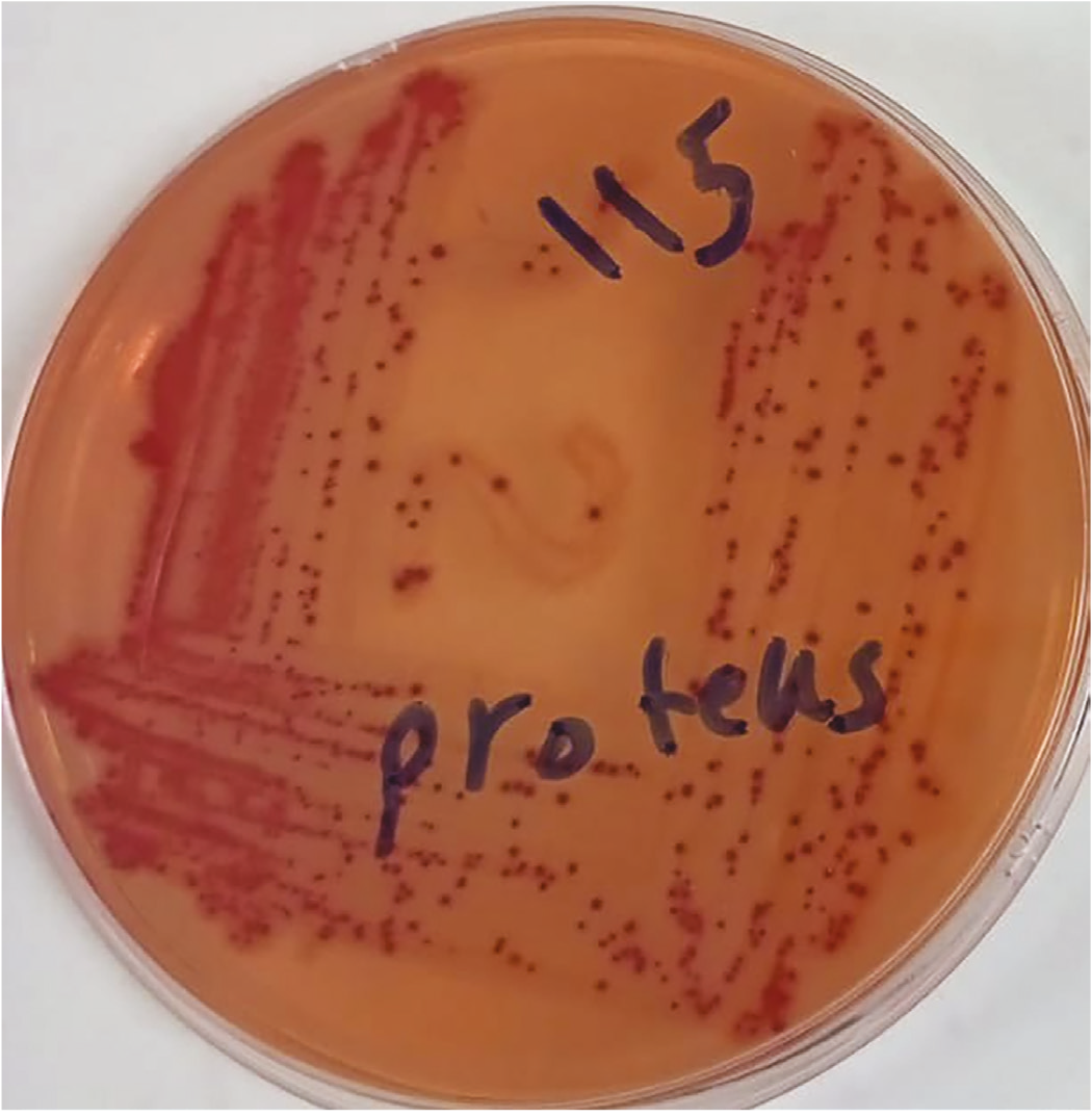
Proteus mirabilis on MacConkey agar.
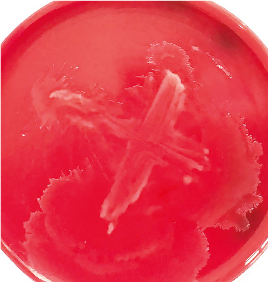
Proteus mirabilis on blood agar.
Table 3. Biochemical test for Proteus mirabilis
Catalase | Oxidase | Citrate | Indole | Urease | MR/VP | TSI |
+ | - | + | - | + | -/+ | H2S+ |
R, methyl red; VP, Voges–Proskauer; TSI, triple sugar iron; H2S, hydrogen sulfide.
The findings of the present investigation suggest that seven (70%) of the Proteus mirabilis isolates had the ureC gene detected by amplifying bacterial DNA using the PCR method at 533 bp, whereas three isolates (30%) were negative (). Moreover, six (60%) of the Proteus mirabilis isolates had the mrpA gene detected by amplifying the bacterial DNA for this gene using the PCR method at 410 bp, while four isolates (40%) did not carry this gene, as shown in .
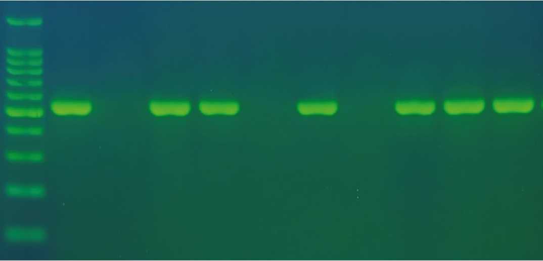
The polymerase chain reaction product exhibits a band size of 533 base pairs. The sample was subjected to electrophoresis on 1.5% agarose using 1 TBE buffer for 1 hour and 30 minutes. N: DNA ladder (100 base pairs).
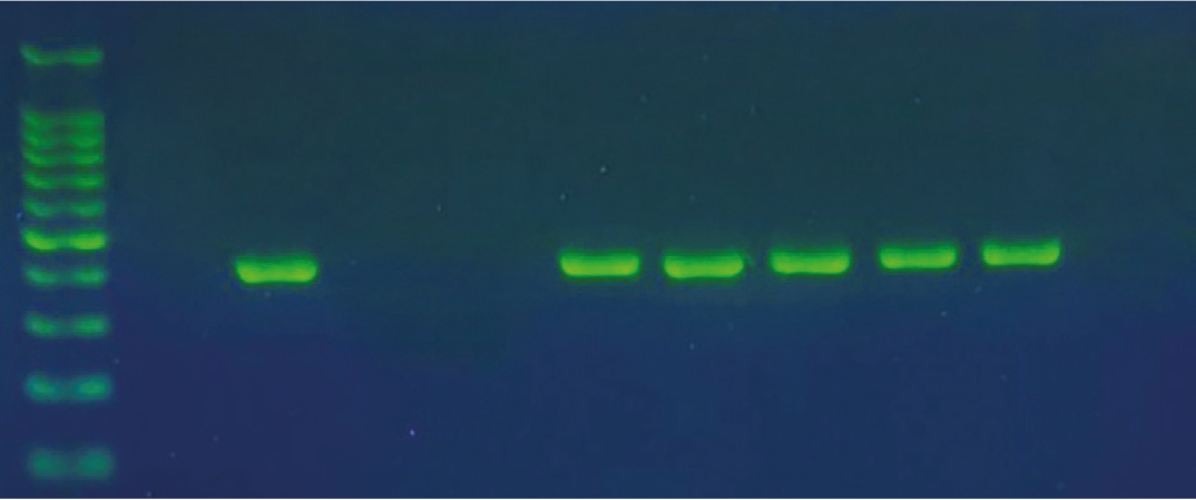
The polymerase chain reaction result shows a band size of 410 base pairs. The sample was subjected to electrophoresis on 1.5% agarose using 1 TBE buffer for 1 hour and 30 minutes. N: DNA ladder (100 base pairs).
In the present study, the mean serum level of IL-6 in the kidney stone patients was 7.62 ± 11.5 ng/mL, while the mean serum level of the control group was 5.05 ± 4.9 ng/mL, as shown in . This result was highly significant (p = 0.002). The mean serum levels of TNF-α and CCL2 in the present study among the kidney stone patients were 18.61 ± 0.21 ng/mL and 211 ± 19.21 ng/mL, respectively, compared with 3.46 ± 0.13 ng/mL and 76.4 ± 5.3 ng/mL in the controls. These differences were also deemed statistically significant (p = 0.001), as shown in and , respectively.
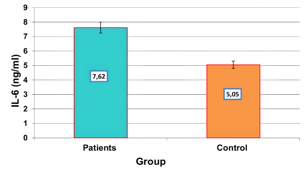
Mean level of IL-6 (ng/mL) in the kidney stone patients and controls. IL-6, interleukin-6.
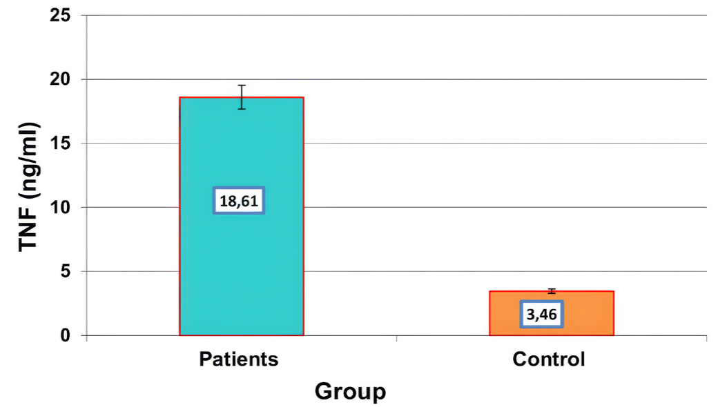
Mean level of TNF-α (ng/mL) in the kidney stone patients and controls. TNF-α, tumor necrosis factor-alpha.
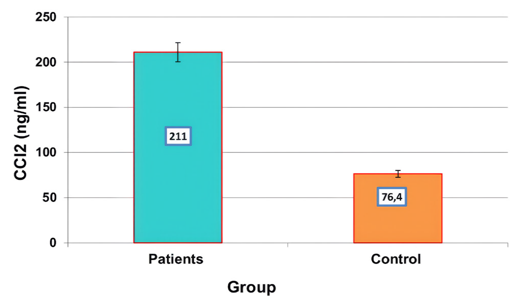
Mean level of CCL2 (ng/mL) in the kidney stone patients and controls.
Discussion
Until recently, it was suggested that only urease-producing bacteria were involved in the pathophysiology of urolithiasis and the formation of struvite stones. Researchers believe that bacteria might help make all kinds of stones, even non-struvite stones. This is because struvite stones are not as common as the other types of stones and people with urolithiasis are more likely to get UTIs and positive urine cultures[23]. The findings of the present study showed that Proteus mirabilis was the predominant bacterium, followed by Escherichia coli and Staphylococcus aureus.
The present study is consistent with previous studies conducted by[24], which reported that the percentage of isolated Proteus mirabilis was 19.04%, and with the study conducted by[25], which indicated that Proteus mirabilis is the most abundant organism in patients with kidney stones. Another study[26] showed a correlation between renal stones and Escherichia coli, Enterococci, Micrococcus luteus, and Staphylococci spp. This result contradicts that of the study conducted by[27], which indicated that Staphylococcus spp. was the predominant genus in the stones and urine of patients with kidney stones. The formation of these outcomes can be attributed to multiple factors, including environmental conditions, host characteristics, and microbial interactions[28]. Antibiotic usage, hospitalization, and host immunological condition may affect the predominance of specific bacterial species. Furthermore, the transmission dynamics of these bacteria in healthcare environments or communities may influence their differing prevalence rates[29]. Proteus mirabilis possesses several virulence genes associated with infection. The results reveal that the prevalence rate of ureC genes was seven (70%) and that of mrpA genes was six (60%). Urease (ureC) is a virulence gene crucial to the pathogenesis of Proteus mirabilis. This enzyme facilitates the formation of kidney stones. It is also crucial for urolithiasis, as it hydrolyzes urea to release ammonia, hence increasing urine pH, which results in the precipitation of calcium and magnesium compounds and the production of kidney stones[12]. The presence of these genes may contribute to the pathogenicity and virulence of the bacterial strains[17]. The mrpA gene, for instance, has been related with certain features, such as adhesion, biofilm formation, and resistance to host defenses[18]. The findings of the present study are consistent with those of a previous study[30] that revealed detection rates of the mrpA gene of 75% but contradict with the findings of[16], which reported a 100% detection rate. The mrpA gene encodes a mannose-resistant Proteus-like fimbriae component needed for urinary tract bacterial adhesion, colonization, and biofilm formation. These fimbriae help bacteria settle in the urinary system and weaken the immune system, prolonging infection. MRPA may increase virulence, which may affect the treatment outcomes by increasing bacterial resistance to human defenses and antimicrobials[31].
The present study indicated an increase in the TNF-α, IL-6, and CCL2 levels in the kidney stone patients compared with the controls. The results are consistent with those of[32-34] indicating increased levels of TNF-α, IL-6, and CCL2. This may result from inflammatory responses induced by the kidney stones, perhaps leading to other urinary system infections. TNF-α is associated with various renal pathologies, and its association with clinical factors has been documented in chronic kidney disease[35]. Ahuja et al.[36] reported that circulating IL-6 drives inflammatory responses through CXCL1 synthesis following acute renal damage in mice. The results indicated that the plasma levels of TNF-α were downregulated, while the plasma concentrations of IL-6 were increased in patients following percutaneous nephrolithotripsy relative to the preoperative values[37]. Boonla et al.[38] showed an increased mRNA expression of IL-6 and MCP-1 in renal biopsy tissue from the kidneys with calculi. The role of IL-6 in stone formation, whether it is released due to the mechanical stimulation of epithelial cells or contributes to renal stone development, remains ambiguous[39,40].
Study Limitations
This study has several limitations that should be addressed. The sample size was relatively limited, and the samples were obtained from a single center, which may affect the generalizability of the findings. The cross-sectional nature of the study does not allow the establishment of causal relationships. However, the use of both microbiological and molecular techniques, along with the evaluation of key inflammatory biomarkers (TNF-α, IL-6, and CCL2), provides valuable insights into the studied condition. Thus, further studies with larger sample sizes and multicenter designs are recommended to further validate these findings.
Conclusion
The present study showed that Proteus mirabilis was the most prevalent bacterium in kidney stone patients. The results indicated that Proteus mirabilis carries the ureC and mrpA genes, which contribute to the pathogenicity of bacteria. Furthermore, this study also suggested that TNF-α, IL-6, and CCL2 tend to increase in kidney stone patients.
Ethics
Ethics Committee Approval: This study was conducted on urine samples collected as part of routine laboratory investigations, with no direct patient intervention. Thus, formal ethical approval was not required according to the institutional guidelines. All procedures were performed in accordance with relevant regulations and ethical standards.
Informed Consent: As the study was conducted using urine samples collected during routine laboratory investigations, without direct patient intervention, the requirement for informed consent was waived in accordance with institutional guidelines.
Ethical Approval
This study was conducted on urine samples collected as part of routine laboratory investigations, with no direct patient intervention. Thus, formal ethical approval was not required according to the institutional guidelines. All procedures were performed in accordance with relevant regulations and ethical standards.
Statistical Analysis
Statistical analyses were performed using the IBM SPSS Statistics software (version 26.0; IBM Corp., Armonk, NY, USA). Data were expressed as mean ± standard deviation. The normality of the data was established using the Shapiro-Wilk test. For the normally distributed continuous variables, the independent samples t-test was used, whereas for the categorical variables, the chi-square test or Fisher’s exact test may be used, depending on the circumstances. Furthermore, statistical significance was set at a p value of <0.05.
Footnotes
Authorship Contributions
Concept: F.A.A.A., A.D.A., Design: A.A.H., Data Collection or Processing: F.A.A.A., A.A.H., A.D.A., Analysis or Interpretation: F.A.A.A., A.A.H., Literature Search: A.A.H., A.D.A., Writing: F.A.A.A., A.D.A.
Conflict of Interest
No conflict of interest was declared by the authors.
Financial Disclosure
The authors declared that this study received no financial support.
References
Elgendy AM, Nabil ZI, Gad El-Hak HN, Nafie MS, El-Shenawy NS. A review of types, mechanisms, complications, and management of renal stone formation. EAJBSC. 2024;16:313-21.
Skolarikos A, Geraghty R, Somani B, Tailly T, Jung H, Neisius A, Petřík A, Kamphuis GM, Davis N, Bezuidenhout C, Lardas M, Gambaro G, Sayer JA, Lombardo R, Tzelves L. European Association of Urology guidelines on the diagnosis and treatment of urolithiasis. Eur Urol. 2025;88:64-75.
Stamatelou K, Goldfarb DS. Epidemiology of kidney stones. Healthcare (Basel). 2023;11:424.
Peerapen P, Thongboonkerd V. Kidney stone prevention. Adv Nutr. 2023;14:555-56.
Xie B, Halter TJ, Borah BM, Nancollas GH. Aggregation of calcium phosphate and oxalate phases in the formation of renal stones. Cryst Growth Des. 2015;15:204-11.
El-Shimy A, Omran A, El-Tair E, Mohamed HI, Kamel I. Identification of different bacterial species isolated from infected renal stones and evaluation of its uricolytic activity. J Med Sci Res. 2018;1:Article 7.
Barbarossa A, Rosato A, Tardugno R, Carrieri A, Corbo F, Limongelli F, Fumarola L, Fracchiolla G, Carocci A. Antibiofilm effects of plant extracts against Staphylococcus aureus. Microorganisms. 2025;13:454.
Kulkarni SR, Peerapur BV, Sailesh KS. Isolation and antibiotic susceptibility pattern of Escherichia coli from urinary tract infections in a tertiary care hospital of North Eastern Karnataka. J Nat Sci Biol Med. 2017;8:176-80.
Wang Z, Zhang Y, Zhang J, Deng Q, Liang H. Recent advances on the mechanisms of kidney stone formation (Review). Int J Mol Med. 2021;48:149.
Sun Y, Wen S, Zhao L, Xia Q, Pan Y, Liu H, Wei C, Chen H, Ge J, Wang H. Association among biofilm formation, virulence gene expression, and antibiotic resistance in Proteus mirabilis isolates from diarrhetic animals in Northeast China. BMC Vet Res. 2020;16:176.
Chakkour M, Hammoud Z, Farhat S, El Roz A, Ezzeddine Z, Ghssein G. Overview of Proteus mirabilis pathogenicity and virulence. Insights into the role of metals. Front Microbiol. 2024;15:1383618.
Hussein EI, Al-Batayneh K, Masadeh MM, Dahadhah FW, Al Zoubi MS, Aljabali AA, Alzoubi KH. Assessment of pathogenic potential, virulent genes profile, and antibiotic susceptibility of Proteus mirabilis from urinary tract infection. Int J Microbiol. 2020;2020:1231807.
Elhoshi M, El-Sherbiny E, Elsheredy A, Aboulela AG. A correlation study between virulence factors and multidrug resistance among clinical isolates of Proteus mirabilis. Braz J Microbiol. 2023;54:1387-97.
El-Tarabili RM, Ahmed EM, Alharbi NK, Alharbi MA, AlRokban AH, Naguib D, Alhag SK, El Feky TM, Ahmed AE, Mahmoud AE. Prevalence, antibiotic profile, virulence determinants, ESBLs, and non-β-lactam encoding genes of MDR Proteus spp. isolated from infected dogs. Front Genet. 2022;13:952689.
Nzakizwanayo J, Pelling H, Milo S, Jones BV. An in vitro bladder model for studying catheter-associated urinary tract infection and associated analysis of biofilms. Methods Mol Biol. 2019;2021:139-58.
Al-Obaidi SA, Al-Hashimy AB. Molecular detection of (ureC, mrpA, hpmA) genes in Proteus mirabilis bacteria isolated from patients with urinary tract infection. Egypt J Hosp Med. 2023;90:716-20.
Aminsharifi A, Molaie A, Monsef A. Laparoscopic ın situ dismembered pyeloplasty can facilitate laparoscopic ureteropelvic junction obstruction repair: a prospective cohort trial. J Endourol. 2018;32:218-22.
Wang B, Tan Z, She W, Wang X, Guan X, Tao Z, Guo F, Xu H, Deng Y. Characterizing chemokine signaling pathways and hub genes in calcium oxalate-induced kidney stone formation: insights from rodent models. Biochem Genet. 2026;64:430-45.
Ni WF, Qin CC. Roles of chemokines in pancreatitis: a review. World J Gastrointest Pathophysiol. 2025;16:111550.
Liu Y, Sun Q, Long H, Qiu Z, Zhang D, Zhang H, Chen J. The value of IL-6, PCT, qSOFA, NEWS, and SIRS to predict septic shock after Percutaneous nephrolithotomy. BMC Urol. 2024;24:116.
Wahid RM, Hassan NH, Samy W, Faragallah EM, El-Malkey NF, Talaat A, Ghoneum A, Aldisi D, Malek MM. The protective effect of allium cepa against ethylene glycol-induced kidney stones in rats. Heliyon. 2023;9:e21221.
Suen JL, Liu CC, Lin YS, Tsai YF, Juo SH, Chou YH. Urinary chemokines/cytokines are elevated in patients with urolithiasis. Urol Res. 2010;38:81-7.
Tavichakorntrakool R, Prasongwattana V, Sungkeeree S, Saisud P, Sribenjalux P, Pimratana C, Bovornpadungkitti S, Sriboonlue P, Thongboonkerd V. Extensive characterizations of bacteria isolated from catheterized urine and stone matrices in patients with nephrolithiasis. Nephrol Dial Transplant. 2012;27:4125-30.
Al-Bassam WW, Al-Kazaz AK. The isolation and characterization of Proteus mirabilis from different clinical samples. J Biotechnol Res Cent. 2013;7:24-30.
Shareef YH, Najem QT, Muhammad JZ. Molecular detection of adhesion genes of Proteus mirabilis isolated from UTI Iraqi patients with kidney stones. MJBU. 2024;42:e185804.
Mahmood AA, Qadir MB, Muhammad AJ, Abdulla TH, Thomas VN, Diyya SMA. Community-acquired infection among the hospitalized renal stone patients. J Health Allied Sci NU. 2024;14:523-6.
Liu F, Zhang N, Jiang P, Zhai Q, Li C, Yu D, Wu Y, Zhang Y, Lv L, Xu X, Feng N. Characteristics of the urinary microbiome in kidney stone patients with hypertension. J Transl Med. 2020;18:130.
Maione A, Galdiero E, Cirillo L, Gambino E, Gallo MA, Sasso FP, Petrillo A, Guida M, Galdiero M. Prevalence, resistance patterns and biofilm production ability of bacterial uropathogens from cases of community-acquired urinary tract infections in South Italy. Pathogens. 2023;12:537.
Nathaniel E, Ikram J, James A, Obaid B, Zahid A, Ahmed Z, Wazir DK, Muhammad Farooq Wahab Q, Varrassi G, Kumar S, Khatri M. Retraction: molecular characterization and antibiotic susceptibility pattern of bacterial strains isolated from wound of patients with diabetes. Cureus. 2024;16:r138.
Hameed Mustafa A. Molecular characterization of multidrug-resistant Proteus mirabilis isolates from pregnant women with recurrent urinary tract infection in Erbil city, Iraq. Iran J Microbiol. 2025;17:751-60.
Li Y, Yin M, Fang C, Fu Y, Dai X, Zeng W, Zhang L. Genetic analysis of resistance and virulence characteristics of clinical multidrug-resistant Proteus mirabilis isolates. Front Cell Infect Microbiol. 2023;13:1229194.
Wang J, Wang W, Guo W, Ma Y, Ji T, Zhang B. Clinical importance of chemokines and inflammatory cytokines for patient care following percutaneous nephrolithotripsy. Exp Ther Med. 2018;15:2189-95.
Xiang L, Zhou D, Zhang H, Li P, Zhou X. Changes of serum and urine cytokines of patients with infectious kidney stones. Chin J Nosocomiol. 2017;27:5172-5.
Sun AY, Hinck B, Cohen BR, Keslar K, Fairchild RL, Monga M. Inflammatory cytokines in the papillary tips and urine of nephrolithiasis patients. J Endourol. 2018;32:236-44.
Budak D, Yilmaz VT, Akbas H, Suleymanlar G, Yucel G. Association between graft function and serum TNF-α, TNFR1 and TNFR2 levels in patients with kidney transplantation. Ren Fail. 2015;37:871-6.
Ahuja N, Andres-Hernando A, Altmann C, Bhargava R, Bacalja J, Webb RG, He Z, Edelstein CL, Faubel S. Circulating IL-6 mediates lung injury via CXCL1 production after acute kidney injury in mice. Am J Physiol Renal Physiol. 2012;303:F864-72.
Kang DH, Rice M, Park NJ, Turner-Henson A, Downs C. Stress and inflammation: a biobehavioral approach for nursing research. West J Nurs Res. 2010;32:730-60.
Boonla C, Hunapathed C, Bovornpadungkitti S, Poonpirome K, Tungsanga K, Sampatanukul P, Tosukhowong P. Messenger RNA expression of monocyte chemoattractant protein-1 and interleukin-6 in stone-containing kidneys. BJU Int. 2008;101:1170-7.
Abd-Alwahab WI, Ajeel AA, Al-Dulaimi FK. The important correlating role of some cytokines with kidney stones in renal calculi patients. AIP Conf Proc. 2022;2394:020012.
Taguchi K, Okada A, Unno R, Hamamoto S, Yasui T. Macrophage function in calcium oxalate kidney stone formation: a systematic review of literature. Front Immunol. 2021;12:673690.